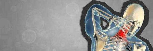
To date, the disease has become very "younger" and is increasingly exposed to people 25 years of age and older, although more recently 30-35 years of age have been considered risky. Cervical pathology is more common, so you should be able to quickly recognize the symptoms of the disease to begin treatment.

So, what is called osteochondrosis of the cervical spine? This term characterizes the degenerative-dystophilic process of the intervertebral disc, which acts as a kind of shock absorber between the segments of the spine. This situation leads to changes in the structure and anatomy, segments, and joint elements of the cervical spine. Osteochondrosis of the neck is characterized by acute pain symptoms that require timely treatment.
Causes of osteochondrosis of the cervical spine
Where does cervical osteochondrosis come from? Listed a little below are the factors whose chronic or acute effects lead to increased neck stress. As a result, the body compensates for the increased loads with the work of the muscles, however, due to the constant tension, cramps occur in them, with damaged blood circulation. Together, these factors lead to degenerative changes in the spine, structural changes, and problems with blood nutrition and metabolism. This is followed by a turn of changes in the intervertebral joints, the bone tissue of the spinal segments overgrowing.
List the factors that contribute to the development of the disease:
- Scoliosis and poor posture.
- Overweight.
- Long stay in a bad and unnatural situation.
- Regular overload of the back and cervical spine, for example, due to the nature of the work.
- Low mobility, sedentary physical inactivity.
- Injury to the spine in the past.
- Metabolism problems.
- Excessive physical activity.
- Stress overload, long-term tendency to depression.
- Inheritance factor.
- Abnormal development of vertebrae.
Grades of cervical osteochondrosis
It is necessary to be able to distinguish between the concepts of "stage" and "degree" that characterize osteochondrosis of the cervical spine. We will examine the stages a little later, and now we will talk about the degrees that depend on the patient’s general clinical condition and complaints, have different symptoms, and require different treatment accordingly.
- First degree - 1. . . Osteochondrosis of the cervix is characterized by minor manifestations of the disease, the main symptoms being pain in the cervical region, which does not appear often, intensifying when you turn your head. They may be accompanied by slightly tense muscles.
- Second degree - 2. . . The severity of the pain and symptoms is much stronger and can give the shoulder area. This is due to the fact that the intervertebral disc became lower in height, leading to nerve stings. Pain syndrome usually increases with movement, and a feeling of weakness and headache leads to decreased performance.
- Third degree - 3. . . This development of cervical spine osteochondrosis is characterized by the development of hernia in the intervertebral space. Deviations from the previous grades occur in even more pronounced and painful symptoms - it gives more shoulder and arm, feeling numb and weak in them. The disease is accompanied by the same headache, weakness, limited neck mobility, and a distinct pain syndrome on palpation.
- Fourth degree - 4. . . This degree is characterized by the complete destruction of the tissues of the intervertebral disc. Problems with the blood supply to the brain are likely to occur primarily through the vertebral artery, which carries blood to the cerebellum and the back of the head. In light of this, coordination, dizziness, ringing in the ear cause difficulties.
Symptoms of osteochondrosis of the cervical spine
Cervical osteochondrosis shows some differences from osteochondrosis in other areas. They arise due to the closer arrangement of the segments relative to each other, the more complex structure of the first two segments - atlas and axis. In addition, there are fewer shock absorbers between the elements of the spine, and accordingly they wear out and degrade faster. In addition, cervical osteochondrosis often leads to compression of the nerves in the spinal cord.
Cervical osteochondrosis - the most common symptoms:
- Painful feelings. . . They are characterized by different localizations - in the back of the head, in the shoulders and in the neck regions. The occurrence of pain in the shoulder joint indicates the pressure of the nerve responsible for transmitting pain impulses at this site. Occipital pain reflects the presence of spasms of the neck muscles in this area due to difficulties in blood flow. Maybe a feeling of pain in his vertebrae, the presence of a crackle.
- Weakness in the hand. . . It is manifested by damage to the nerve responsible for motor activity in the upper extremities.
- Weak sensitivity in hand. . . The nerve that innervates the skin of the arm is damaged.
- Restricted movement, crunching. . . This is due to the low height of the intervertebral disc, the growth of bone in the segments of the spine, and the presence of small affected structures.
- Coordination problems, weakness and dizziness. . . As the pathology progresses, fibrous tissue is formed. In part, it leads to a contraction of the vertebral artery, which has its own channel in the elements of the spine. This reduces the lumen of the blood vessel, causing blood deficiency in the occiputum and cerebellum.
- Hearing, vision, speech problems. . . These further develop the contraction of the cerebellum and the vessel feeding the occipital zone.
Diagnostics
Diagnosis is made in the presence of a person’s characteristic symptoms and complaints. Osteochondrosis of the cervical spine is diagnosed by various methods, primarily to visualize the condition of the injured part. Most commonly used:
- Radiography. It is not very informative, it only shows the presence of abnormalities, it is mainly suitable for early diagnosis.
- Computed tomography. Compared to radiography, the visualization of pathologies of the cervical spine segments is improved, but this does not accurately determine the presence of the hernia and its size. Furthermore, this procedure cannot determine the "narrowing" of the channel with the spinal cord.
- Magnetic resonance imaging. Such a diagnosis is characterized by state-of-the-art, increased information content, allowing a detailed assessment of bone structure defects, condition of intervertebral discs, existence, size, and direction of growth of hernias.
- If deterioration in the flow of vertebral arteries is suspected, further diagnosis will be made using the ultrasound duplex scanning procedure. Such a study accurately identifies the presence of barriers that reduce the rate of blood flow.
Based on the data obtained during the diagnosis, we can talk about different stages:
- Section 1, characterized by a minor violation of the anatomy of the vertebrae.
- Section 2. . . It is likely that ignoring the relative position of the vertebrae, displacement, rotation relative to the axis of the spine, the intervertebral disc may be slightly reduced in height.
- Section 3. . . The height of the disc decreases by a quarter, the joints change, there are outgrowths of the bone tissue, the tension of the intervertebral foramen and the spinal canal.
- Section 4. . . It still got worse than the previous one. The height of the disc is greatly reduced, with deep joint pathologies and extensive bone growths behind it, the ducts of the spinal canal and the spinal cord are strongly compressed.
Treatment of osteochondrosis of the cervical spine
The main methods of such treatment: drug therapy, physiotherapy, the use of massage in the affected area, therapeutic gymnastics. Let’s take a closer look at some of the methods.
Drug treatment
Any medication should only be prescribed by a qualified professional.
- Non-steroidal anti-inflammatory drugs. Their effect is pain syndrome, effective removal of the inflammatory and edematous process of compressed nerve endings.
- Vitamin B is taken to improve the metabolic processes in the vertebrae and nerves.
- Drugs that increase blood flow. It is used to nourish altered nerve endings and improve blood flow to the brain.
- Chondroprotectors for cartilage and intervertebral disc tissue repair.
- Muscle relaxants, antispasmodics.
Physiotherapy
- Electrophoresis. . . Delivery of drug ions to the desired part of the pathology by an electric field. Before the procedure, novocaine anesthesia is performed and aminophylline is also used, which improves blood flow.
- Ultrasound. . . Relieves inflammation, pain, promotes metabolism at the site of application.
- Magnetotherapy. . . It has an analgesic effect, relieves swelling.
- Laser therapy. . . The treatment is exposed to light waves of special frequencies. It relieves inflammation well and promotes blood circulation.
Physiotherapy
Physiotherapy is allowed only in the absence of exacerbation of the disease. The techniques will be effective in the absence of pain and discomfort during implementation, and will be very effective as a means of prevention. Here are some basic exercises:
- Lie on your stomach and support the bent arms on the floor. Perform head and torso lifts for 60-90 seconds, keep your back straight, and then return smoothly to its original position. Perform 2-3 repetitions.
- Lie on your stomach, arms outstretched along the torso. Turn your head left, right while trying to reach the floor with your ears. Perform 5-7 repetitions on each side.
- In a sitting position, inhale, lean forward, trying to reach the chest with your head. Then, when exhaling, on the contrary, sit back, throw your head back. Perform 12 repetitions.
- In a sitting position, place your palm on your forehead. Apply mutual pressure on the forehead on the palm and vice versa. Continue for up to half a minute, repeat three times.
- Unhurried, lateral rotation of the heads, 5-7 rotations each. If you feel dizzy, reduce the number of repetitions or slow down.
Cervical spine osteochondrosis: question-answer
Does osteochondrosis of the cervical spine need to be treated with surgery?
It happens, but in the rare cases where conservative treatment does not produce an effect half a year after the start. In such situations, there is usually chronic pain and tension in the nerve endings.
How long can you take non-steroidal anti-inflammatory drugs?
Most of these drugs affect the condition of the gastric mucosa, so their frequent and regular use is not recommended. The exact duration of medication is determined by the treating physician, who has information about the stage of the patient's osteochondrosis and whether there are concomitant pathologies.
Are chondroprotective drugs effective in repairing damaged intervertebral disc tissue?
The potency of such drugs has not been proven, so they are rarely recommended by doctors.
What should we do at home if there is no aggravation in order to avoid aggravation in the future?
For prevention purposes, best physiotherapy practices, use of cervical zone massage, rejection of significant physical activity.


